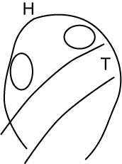
These images were obtained using a Meiji 1-7x zoom microscope and a Sony DSC-F707 5 Mega-pixel camera. The images of eggs are at 7x, fish at 1x. The "Day numbers" are educated guesses at best. Eggs were being laid during the period of collection, and the temperature, which varied a great deal, has a big effect on development rate.
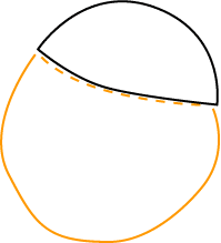
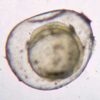
Day 1: Cells have been rapidly dividing since fertilization of the egg; they form a cap (indicated by the black line) over the yoke mass (yellow line). The yellow line is dashed where the yoke extends up under the cell cap.
The egg is about 1 mm in diameter.
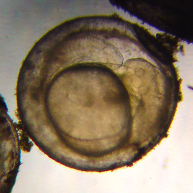
Day 3: Cells have divided and migrated to form a tube (the spinal cord) with bulges at the two ends.
Day 5: The differential cell division and migration has created a much more elongated tube which now stretches around more than half of the yoke sac.
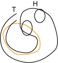
Day 7: Now the form of the adult fish is evident because the eye can be seen (the small circle in the head). The fish makes one turn around the yoke sac.
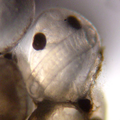
Day 9: The body has become larger, but the dramatic change is the production of black pigment in the eyes.
Day 10: The head has grown so large that it appears to fill the egg. The body (mostly tail) is coiled inside the egg shell for several turns around the yoke sac. You can only see part of the tail here.
Day 10 plus: Now you can see how crowded it was in the egg, am I glad to be free!
The "stomach" is what's left of the yoke sac, which will sustain the fry until it starts to eat. This fry is about 11 mm long, while the egg was the size of the orange circle, 1.2 mm in diameter. Thus, inside the egg the tail was coiled more than twice around the yolk.