Physiology
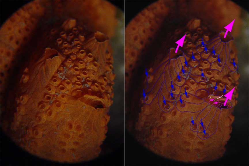
Figure B-1. Water flow through a colony.
Each zooid of a colony has an intake vent, but as they merge to make a colony they lose their outer walls and water is discharged from large common exit vents.
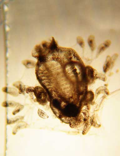
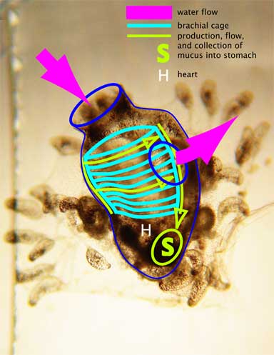
Figure B-2. How a zooid eats.
This zooid has recently developed from a “tadpole”. It’s about 1 mm across and has attached to a microscope slide. Water is pumped through the zooid by thousands of beating cilia, too small to be seen here, and is forced through a mucoid net stretched around the inside of the brachial cage. The net is continually produced by the endostyle, a linear array of mucoid producing cells along the side of the cage opposite the exit vent. Along the side of the cage opposite the endostyle, the net is coiled and pulled down into the stomach where the net and all trapped organisms and food fragments are digested.
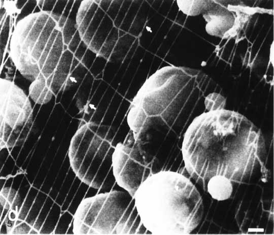
Figure B-3. The mucoid feeding net.
The net is very fine, and can’t be resolved by my modest equipment. This image of a net produced by another tunicate was obtained using a scanning electron microscope (SEM), and appears in the book “The Biology of Pelagic Tunicates”, edited by Q. Bone. The small white bar in the lower right corner represents 1 micron (micrometer). Bacterial are typically 1-3 microns in diameter, and the wavelength of light is about 0.5 microns. The net is similar to a spider web, except it has rectangular, not radial symmetry. The tunicate net is fabricated by movements of a linear array of cilia, not by movement of the entire animal, as a spider builds its web.


